


Website by ![]()
Our Blog
The Role of Ultrasound in Detecting Vascular Blockages
At GLMI, we offer advanced vascular ultrasound testing to help patients across Buffalo, Williamsville, Cheektowaga, Orchard Park, and Cambria identify circulation problems early. This non-invasive imaging technique plays a crucial role in diagnosing arterial and venous blockages — helping physicians develop timely and targeted treatment plans.
What Is a Vascular Ultrasound?
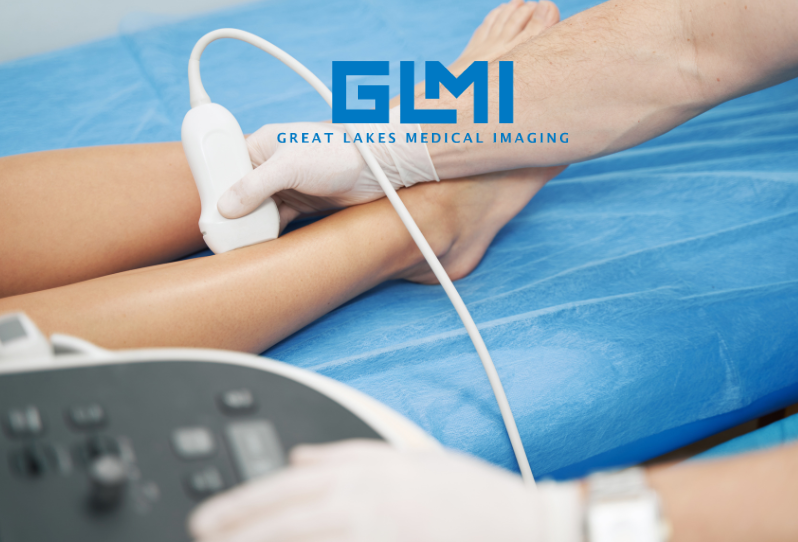
A vascular ultrasound, sometimes called a Doppler ultrasound, uses high-frequency sound waves to create real-time images of blood vessels and measure blood flow. Unlike X-rays or CT scans, ultrasound does not use radiation, making it a safe and comfortable option for patients of all ages.
During the exam, a technician applies a small amount of gel to the skin and moves a handheld device (called a transducer) across the area being studied. The sound waves bounce off blood cells and vessel walls, creating detailed images on a monitor. Doppler technology allows clinicians to see not only the structure of the vessels but also how well blood is flowing through them.
Why Ultrasound Is Ideal for Detecting Vascular Blockages
Ultrasound offers a clear and accurate view of both arteries and veins, allowing doctors to detect even minor abnormalities in blood flow. It’s one of the most valuable diagnostic tools for identifying:
- Arterial narrowing (stenosis): Caused by plaque buildup inside arteries, which restricts blood flow and can lead to peripheral artery disease (PAD) or stroke.
- Venous insufficiency: When vein valves fail to function properly, leading to pooling blood, swelling, and varicose veins.
- Blood clots (deep vein thrombosis): Ultrasound can reveal the presence of clots that could dislodge and travel to the lungs.
- Aneurysms: Weak spots in blood vessel walls that can balloon and potentially rupture.
- Bypass grafts or stent evaluations: Ultrasound helps monitor the success and long-term function of vascular procedures.
Because it captures blood flow in real time, ultrasound provides immediate insights into how well your circulatory system is performing — without invasive testing or exposure to contrast dye.
Common Types of Vascular Ultrasound Studies
GLMI offers several types of vascular ultrasound exams, depending on which part of the body needs evaluation. Each test serves a unique purpose in detecting blockages and assessing overall vascular health.
1. Carotid Ultrasound
This test evaluates the carotid arteries in the neck, which supply blood to the brain. It’s used to detect narrowing or plaque buildup that can increase the risk of stroke. Early detection allows for lifestyle changes or medical intervention before serious complications occur.
2. Peripheral Arterial Ultrasound
This study examines blood flow in the legs and feet. It’s commonly used to diagnose peripheral artery disease (PAD), a condition that causes leg pain, cramping, or fatigue due to restricted circulation. Early detection through ultrasound helps prevent ulcers, infections, and tissue damage.
3. Venous Ultrasound
Venous ultrasound evaluates the veins that return blood to the heart. It’s used to detect deep vein thrombosis (DVT) and assess venous insufficiency. It can also identify causes of leg swelling or varicose veins.
4. Abdominal Aortic Ultrasound
This test checks for aneurysms (abnormal bulges) in the abdominal aorta — the main artery that carries blood from the heart to the rest of the body. Detecting aneurysms early can prevent potentially life-threatening ruptures.
5. Upper Extremity Ultrasound
Used to evaluate blood flow in the arms, this test can identify vascular blockages or clots related to repetitive strain, trauma, or medical device placement.
Symptoms That May Indicate a Vascular Blockage
Because many vascular problems develop gradually, early warning signs can be easy to overlook. You should talk with your doctor about a vascular ultrasound if you experience any of the following:
- Cold, pale, or discolored feet or toes
- Cramping, numbness, or weakness in the legs during activity
- Swelling or heaviness in the legs or ankles
- Visible varicose or spider veins
- Non-healing wounds or ulcers on the lower legs
- Shortness of breath or chest pain (possible sign of a related circulatory issue)
- Dizziness or sudden vision changes (possible sign of carotid artery narrowing)
Even if symptoms are mild, ultrasound can help determine whether reduced blood flow is contributing to your discomfort.
Advantages of Ultrasound for Vascular Diagnosis
There are many reasons why ultrasound is a first-line imaging tool for detecting vascular blockages:
- Non-invasive: No needles, injections, or surgical procedures are required.
- Safe: Ultrasound uses sound waves, not radiation.
- Immediate results: Technologists and radiologists can quickly evaluate images for signs of restricted flow.
- Affordable and accessible: It’s widely available and cost-effective compared to other imaging methods.
- Highly accurate: Modern Doppler ultrasound systems provide precise measurements of blood flow velocity and vessel integrity.
Because it combines accuracy with comfort, ultrasound is often the preferred diagnostic choice for vascular evaluation — and a vital tool for early detection and prevention.
How GLMI Uses Ultrasound to Improve Vascular Health
At GLMI, our vascular imaging program is designed to help physicians detect blockages and circulation problems as early as possible. Our board-certified radiologists and experienced technologists use advanced ultrasound systems to evaluate blood flow in real time, ensuring accurate results and a comfortable patient experience.
We work closely with primary care doctors, cardiologists, and vascular specialists to interpret findings and develop personalized care plans. Whether you’re being screened for venous disease, evaluated for peripheral artery disease (PAD), or monitoring post-surgical results, our team delivers clear answers that guide better outcomes.
Why Early Detection Matters
Vascular blockages can lead to severe health issues if left untreated, including heart attack, stroke, or limb-threatening circulation problems. Early diagnosis through ultrasound allows your doctor to recommend treatment strategies such as lifestyle changes, medications, or minimally invasive vascular procedures to restore blood flow and prevent complications.
By combining preventive imaging with healthy habits — such as regular exercise, balanced nutrition, and smoking cessation — patients can significantly reduce their risk of developing advanced vascular disease.
Why Western New York Patients Trust GLMI
GLMI has been a leader in diagnostic imaging and vascular care for decades. Our ultrasound services and vein and vascular programs provide Western New York patients with accurate results, fast appointments, and compassionate care close to home. With locations in Williamsville, Cheektowaga, Orchard Park, and Cambria, we make advanced vascular testing convenient and accessible.
Schedule a Vascular Ultrasound Today
Don’t wait for circulation problems to worsen. Early testing through vascular ultrasound can identify blockages before they lead to more serious health concerns. If you’re experiencing symptoms of poor circulation or have risk factors for vascular disease, imaging can help you take control of your health today.
Contact GLMI to schedule your vascular ultrasound appointment and learn more about our advanced diagnostic services in Western New York.
Medical Disclaimer
Medical Disclaimer: This article is for informational purposes only and is not a substitute for professional medical advice. Always consult your physician or qualified healthcare provider about your specific symptoms and treatment options.
‹ Back