



Website by 
Our Blog
Wrist and Elbow Injuries: Imaging Sprains, Fractures and Tennis Elbow
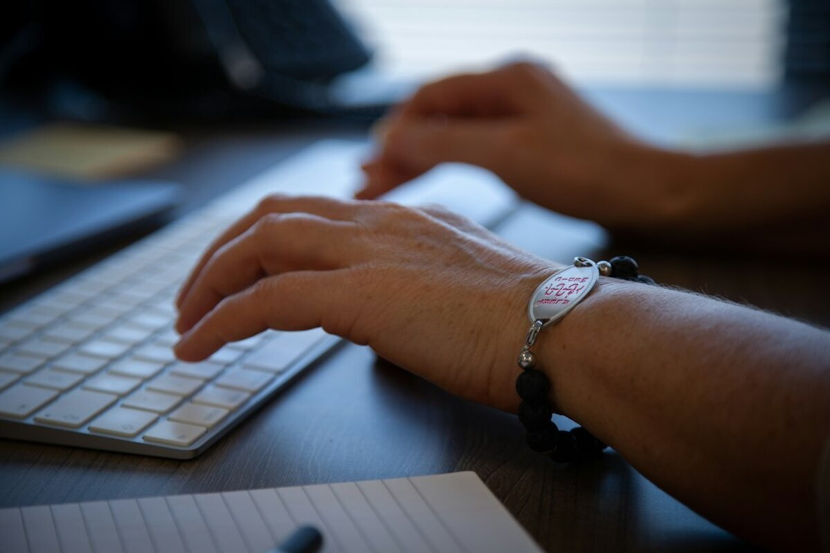
You slipped on black ice at the end of the driveway, threw a hand down to catch yourself, and got up more embarrassed than hurt. That was ten days ago. The swelling went down. But you still cannot turn a doorknob without wincing, and holding a coffee cup on that side feels wrong in a way you cannot explain.
Wrists and elbows are the overlooked joints. Knees and shoulders get the braces and the sideline drama. Wrists and elbows just quietly stop working, and because their injuries look unremarkable from the outside, people wait. Sometimes that is fine. Sometimes it turns a small problem into a much harder recovery. Here is how to tell the difference.
The Fall on an Outstretched Hand
Orthopedic offices have shorthand for this one: FOOSH, short for fall on an outstretched hand. It is the most common way people around Buffalo hurt a wrist between December and March, and just as common on a soccer field or on the back steps.
The force travels up through your palm into the two forearm bones, and the radius, the thicker bone on the thumb side, absorbs most of it. A distal radius fracture, meaning a break near the wrist, is a frequent result. When the break is displaced, the wrist looks visibly crooked and nobody has to talk you into getting it checked. The tricky ones are non displaced fractures, where the bone cracked but the pieces stayed in line. Those look almost normal. You can still move the wrist, type, and drive.
An X-ray is the right first test, and it answers the bone question fast. If the images look clean and your wrist still hurts a week later, that is not the end of the conversation. It is the start of the next one.
The Scaphoid Fracture That Hides
One small bone deserves its own section. The scaphoid sits at the base of your thumb, under the soft hollow you can feel when you stick your thumb out sideways. Anatomists call it the anatomic snuffbox. Press there after a fall, and if it is sharply tender, pay attention.
Scaphoid fractures are notorious for two reasons. First, they often do not show up on the initial X-ray. The crack can be so fine, and the bone so oddly shaped, that the fracture line hides behind overlapping bone on day one. Second, the scaphoid has an unusual blood supply that enters near the far end and flows backward through the bone. A fracture across the middle can cut off circulation to the piece closest to the forearm, so the bone may fail to heal or the fragment may slowly die. Both are much harder to fix than a fracture caught early.
This is why a careful clinician will not simply say "the X-ray is negative, it is a sprain." The usual approach is to splint the wrist and either repeat the X-ray a week or two later, once healing changes make the line visible, or go straight to MRI, which sees swelling inside the bone within days of the injury. Being told to come back for a second set of images is not indecision. It is respect for a bone with a bad reputation.
When a Sprain Is Really a Torn Ligament
"Sprain" is a broad word. It covers a ligament that will be fine in a week and a torn one that changes how the wrist bones move. Two tears in particular get missed. The scapholunate ligament keeps the small wrist bones tracking together, and when it tears they drift out of position, which over years wears the cartilage down. The second is the triangular fibrocartilage complex, or TFCC, a cushion and sling of tissue on the pinky side of the wrist. TFCC injuries hurt along that outer edge, often with a click, and they announce themselves when you twist: turning a key, opening a stubborn jar, pushing up out of a chair. Neither shows on a plain X-ray, because ligaments and cartilage are soft tissue and X-ray is built for bone. MRI answers that question.
Tennis Elbow and Golfer's Elbow Are Tendon Problems
Move up the arm and the story changes. Most elbow pain in adults is not a joint problem. It is a tendon problem at one of the bony bumps on either side.
Tennis elbow, or lateral epicondylitis, affects the tendon anchoring your wrist extensor muscles to the outside of the elbow. Golfer's elbow, medial epicondylitis, affects the flexor tendon on the inside. Despite the names, most people who get these have never held a racquet or a seven iron. Painters, electricians, and anyone who spent a weekend gripping a power tool are just as likely. And despite the "itis" ending, which implies inflammation, what is usually happening is tendinosis, meaning the collagen fibers have degenerated from repetitive load. That distinction matters, because the two respond to different treatments.
These tendons sit close to the skin, which makes them ideal for diagnostic ultrasound. Ultrasound shows the tendon in high resolution, in real time, while you move the elbow, which no other test can do. A radiologist can see whether it is thickened, whether there are darker regions where the fiber structure has broken down, and whether part of the tendon has pulled off the bone. Doppler, the setting that maps blood flow, reveals the abnormal small vessels that grow into a chronically irritated tendon. No radiation, no contrast injection.
If elbow pain has outlasted a few weeks of rest and a brace, ultrasound can tell your physician whether they are managing an irritated but intact tendon or a partial tear. Call GLMI at 716-836-4646 or use our scheduling information page to ask about a musculoskeletal ultrasound, and bring your referral so we can often get you in the same day.
Throwers and the Ulnar Collateral Ligament
Baseball and softball players have a different elbow problem. Every throw puts enormous stretching force on the inside of the elbow, and the ulnar collateral ligament is what resists it. Pitchers who are overworked, or who ramp up too fast in spring after a Western New York winter spent indoors, can strain or tear it.
The warning signs are specific. Pain on the inner elbow during the acceleration phase of the throw. A drop in velocity or control the athlete cannot explain. Tingling in the ring and little fingers, because the ulnar nerve runs right behind that ligament. MRI is the standard test, because it grades the ligament from mildly strained to fully torn, and that grade shapes whether the plan is rest and a throwing program or a surgical consult. GLMI is the official imaging provider for the Buffalo Bills, and the same sports injury imaging we perform for professional athletes is available to a high school pitcher in Cheektowaga.
When to Stop Icing and Get Imaged
Ice and a few days of patience are reasonable for most minor injuries. Here is when that plan has run out of road.
- Sharp tenderness in the anatomic snuffbox after a fall, even if an X-ray was read as normal
- Pain or swelling that has not clearly improved after ten to fourteen days
- You cannot grip, rotate your palm up and down, or fully straighten the elbow
- Visible deformity, or numbness and tingling in the hand
- A pop or snap at the moment of injury, especially in a throwing athlete
- Pain that wakes you at night or returns every time you go back to activity
What an Appointment Looks Like
At GLMI, wrist and elbow questions usually start with X-ray. From there, ultrasound handles the tendons and MRI handles the ligaments, cartilage, and hidden fractures. If lying still inside a scanner is a problem for you, ask about our OPEN 1.2T High Field magnet, built for people who find enclosed spaces difficult. Every study is read by a fellowship trained radiologist, and the report goes to your referring physician so decisions do not stall. None of this replaces an exam, but better pictures make for better decisions.
If your wrist or elbow still hurts after a couple of weeks and nobody has looked past that first X-ray, this is the week to change that. Call 716-836-4646 or reach us through our contact page to arrange imaging in Williamsville, Cheektowaga, Orchard Park, or Cambria, and let your doctor work from a clear picture instead of a guess.
‹ Back
Recent Posts
-
-

-

Breast Biopsy: What to Expect, Step by Step
October 16, 2026
-
High School Football Injuries: When Your Athlete Needs Imaging
October 13, 2026
-

Breast MRI: Who It Is For and What It Shows
October 10, 2026