



Website by 
Our Blog
High School Football Injuries: When Your Athlete Needs Imaging
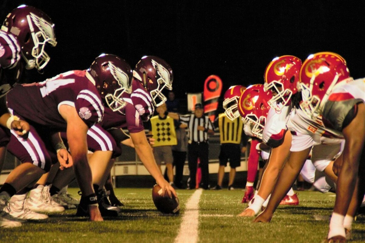
The metal bleachers are about forty degrees, you have a blanket over your knees and a thermos going cold beside you, and your kid just got rolled up on near the far sideline. He limps off under his own power. The trainer looks at the ankle, the coach says ice it tonight, and somewhere between the parking lot and your driveway in Hamburg you start wondering what happens Monday if it still hurts.
High school football in Western New York packs a lot of contact into a short season, and parents end up making quick decisions with incomplete information. Here is a plain guide to the injuries that send teenagers for imaging, what each scan can and cannot tell you, and how to tell sore from hurt.
Start With the Question, Not the Machine
Families sometimes arrive asking for an MRI the way you would ask for a specific tool at a hardware store. It is a reasonable instinct, but imaging works in reverse. A physician or athletic trainer examines the joint, forms a short list of what could be wrong, and picks the study that separates those possibilities. An MRI ordered without a question produces findings that sound alarming and mean little, especially in a growing athlete whose joints are changing anyway.
Shoulders: Separations, Dislocations and Cuffs
Shoulders take a beating because players land on them and drive into contact, and two injuries get confused constantly. A separation involves the AC joint, where the collarbone meets the top of the shoulder blade, and it follows a hard landing on the point of the shoulder. A dislocation is the ball of the upper arm leaving the socket entirely.
X-ray is the starting point for both. It shows bone alignment, whether the collarbone has ridden up, and whether anything is fractured. When pain persists after a clean bone picture, or an arm keeps slipping out, MRI takes over. It shows the labrum, the rim of cartilage that deepens the socket, and the rotator cuff, the four muscles and tendons that hold the ball centered and lift the arm overhead.
Full cuff tears are mostly an adult injury. In teenagers, lingering shoulder pain is more often a strained cuff, an irritated growth area, or instability from a labral injury, and MRI tells those apart.
Knees: The One Every Parent Dreads
You can often guess a knee injury from the description. A non-contact plant and pivot, a pop, and swelling that balloons within an hour points toward the ACL, the ligament through the center of the knee that keeps the shin bone from sliding forward. A hit from the outside that bends the knee inward tends to strain the MCL along the inner side. Twisting with the foot planted, then clicking, catching or a knee that will not fully straighten, suggests the meniscus, the two C shaped cushions of cartilage between the bones.
Ligaments and cartilage are soft tissue, and soft tissue is MRI territory. X-ray still matters at the first visit, since it rules out a fracture and shows whether the injury pulled a piece of bone away from a growth area. But the study that names the injury, grades it, and gets looked at before anyone discusses surgery is an MRI. Our 1.5T High Field MRI handles knees beautifully, and for athletes who dislike enclosed spaces we also run a wide bore 3.0T and an open magnet.
Ankles, and What Hides Behind a Sprain
Ankles roll constantly, and most of those are ligament sprains that settle down with rehab. The ones worth a closer look have tells: pain directly over the bony knobs rather than the soft tissue below them, an inability to bear weight for more than a couple of steps, a foot that looks off axis, or pain high above the ankle between the two shin bones, which can mean a high ankle sprain and a much longer timeline.
X-ray answers the fracture question quickly. When the bone looks fine and pain is not improving on schedule, ultrasound or MRI shows the ligaments and tendons directly. Ultrasound has a real advantage for some tendon problems, since we can watch the tendon move while you flex the joint, with no radiation.
Growth Plates: The Part That Is Different in Teenagers
Most parents have never had this part explained, and it matters more than the rest. A growth plate is a layer of cartilage near the end of a long bone where new bone is added while a kid is growing. That cartilage is softer than the bone around it and softer than the nearby ligaments.
In an adult, a violent twist tears a ligament. In a fourteen year old, the same force can pull through the growth plate instead, because it is the weakest link in the chain. What a coach would call a bad sprain in a grown man can be a growth plate fracture in a teenager, and some are subtle on a first X-ray. Two related patterns show up in football too. Apophysitis is irritation where a big tendon attaches to a growing area, including just below the kneecap and around the pelvis. An avulsion is when a hard sprint or kick yanks that attachment loose along with a fragment of bone.
The takeaway is simple. Teenage pain that sits right on a bone, close to a joint, and does not improve over several days deserves a real evaluation, not another week of ice.
If your athlete's doctor has sent you out for imaging and you do not want to lose a week to scheduling, call 716-836-4646 or use our scheduling information page. Same-day appointments are often available at our Williamsville, Cheektowaga, Orchard Park and Cambria locations, and we can frequently handle an X-ray and an MRI in one trip instead of two.
Stingers, and When a Nerve Injury Is Not Minor
A stinger is that lightning bolt of burning pain down one arm after a collision, usually with numbness or a dead arm for a minute or two. The nerves running from the neck into the arm get stretched or compressed, and most stingers clear quickly enough that the player goes back in.
The ones that need attention break that pattern. Symptoms in both arms, weakness that hangs around after the burning stops, midline neck pain, a stinger lasting more than a few minutes, or an athlete collecting them repeatedly all point toward getting the neck evaluated. That usually starts with X-ray of the cervical spine and moves to MRI if there is concern about the discs, the spinal cord or the nerve roots.
Matching the Scan to the Question
If you remember nothing else, remember this:
- X-ray for bone: fractures, alignment, dislocations, and a first look at growth plates. Usually the first study ordered.
- MRI for soft tissue: ligaments, cartilage, tendons, muscle, discs, and bone bruises that X-ray cannot see.
- Ultrasound for certain tendon and muscle questions, with live motion and no radiation.
Head injuries follow their own rules and belong with your physician or an emergency department rather than a scheduled outpatient scan.
The Same People Who Read for the Bills
Great Lakes Medical Imaging is the official imaging provider for the Buffalo Bills, so the fellowship-trained radiologists reading your son's knee MRI are the same team trusted with professional athletes. A junior in Orchard Park gets the same quality of read, and that matters when a sports medicine physician is deciding off the report.
Speed matters too. A high school season is short, and an athlete waiting in limbo for a scan is losing time he cannot get back. Our sports injury imaging service is built around that, with same-day appointments and quick turnaround so treatment can start while the season is still going.
When Monday Morning Comes
Most Friday night injuries are what they look like, and a weekend of ice and rest sorts them out. The ones that are not announce themselves: they refuse to improve, they swell out of proportion, the joint gives way or locks, or the pain sits on bone rather than in muscle. Trust that pattern more than your teenager's insistence that he is fine.
When the answer is not obvious, we can help you get it fast. Text our sports injury line at 716-333-7773, call 716-836-4646, or reach us through the contact page, and we will get the right scan scheduled so your athlete can start the road back.
‹ Back
Recent Posts
-
High School Football Injuries: When Your Athlete Needs Imaging
October 13, 2026
-

Breast MRI: Who It Is For and What It Shows
October 10, 2026
-
Concussion Imaging: When a Head Injury Needs a Scan
October 7, 2026
-

Called Back After a Mammogram? What It Does and Does Not Mean
October 4, 2026
-