



Website by 
Our Blog
Concussion Imaging: When a Head Injury Needs a Scan
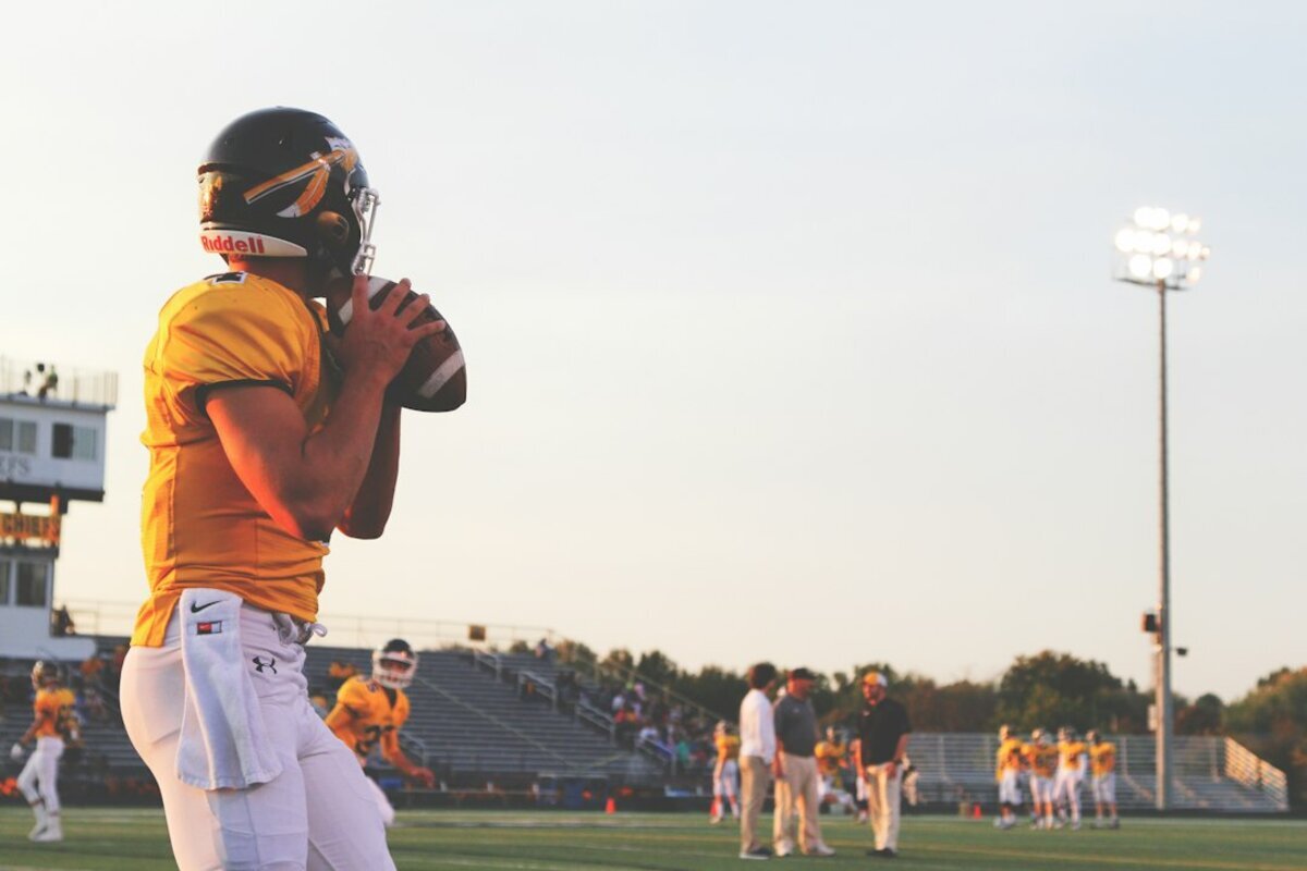
It is nine o'clock on a Sunday night in October. Your son took a helmet to the side of the head in the third quarter, sat out the rest of the game, and has been unusually quiet since. He says he is fine. He has also asked you twice what time school starts. You are standing in the kitchen with your phone in your hand, deciding whether this is an ice pack and an early bedtime or a drive to the emergency room.
Families all over Western New York run this same calculation, and not only during football season. A slip on an icy driveway in Amherst produces the identical worry in January. What almost everyone wants to know is whether a scan would settle it. The honest answer starts with something that surprises most people.
A Concussion Does Not Actually Show Up on a Scan
This is the part that catches families off guard. A concussion is a functional injury, not a structural one. The brain moves suddenly inside the skull, and the cells briefly stop handling energy and chemical signaling the way they normally do. The wiring is stressed on a microscopic level. Nothing breaks in a way a camera can photograph.
CT and MRI scanners are structure machines. They show anatomy: bone, blood, fluid, tissue that has shifted out of place. So a person can have a textbook concussion, with headache, fog, light sensitivity and a short temper for two weeks, and have a completely normal scan. That does not mean the injury was imagined. It means the injury lives at a scale the scanner cannot see. So if the scan cannot find the concussion, what is it looking for?
The Scan Is Hunting the Things That Get Worse
Imaging after a head injury is not there to confirm the concussion. It is there to rule out the small number of injuries that can turn dangerous in the hours after a hit, often while the person is asleep and no one is watching.
The main ones are bleeding inside or around the brain, a skull fracture, a bruise on the brain tissue itself, and swelling. Bleeding drives most emergency scans. When a vessel tears, blood collects in a space that has nowhere to expand, because the skull is a sealed box. Pressure builds. The person may look fine for an hour and then decline quickly. That window is why the decision about imaging gets made tonight rather than in the morning.
A clean scan is good news. It says the emergency version of this injury is not happening. It does not say you can stop paying attention.
The Signs That Change the Answer
Doctors lean on a handful of warning signs when deciding who needs immediate imaging. If any of these are present, the situation belongs in an emergency room right away, not in a scheduling queue:
- A headache that keeps getting worse instead of settling down
- Vomiting more than once
- Confusion, not recognizing familiar people or places, or repeating the same question
- Any loss of consciousness, even brief
- Pupils of different sizes, double vision, or slurred speech
- Weakness, numbness or clumsiness on one side of the body
- A seizure, or clear fluid or blood coming from the nose or ears
- Someone who cannot be woken normally, or is becoming harder to rouse
- A blood thinner in the picture, which we will come back to
Notice how many are about direction rather than intensity. A moderate headache that is slowly fading is a different animal from one that has been climbing for three hours. Trajectory is the thing to watch.
One practical note. Emergency head imaging happens at a hospital, because that is where the surgical and neurological help sits if the scan finds something. Great Lakes Medical Imaging is an outpatient practice, so we handle the planned imaging your physician orders before or after that acute moment.
When Watching and Waiting Is the Right Call
Most head injuries do not need a scan at all, and a clinician who tells you to go home and monitor is not brushing you off. With no warning signs, a person who was oriented and coherent shortly after the hit, and symptoms holding steady or improving, careful observation is a legitimate plan rather than a consolation prize.
Imaging carries its own considerations, particularly in children and teenagers, so the goal is to scan the people who need it and spare those who do not. Your physician will tell you what to watch for over the next day or two and when to stop waiting. Keep that list where the household can see it, because the person with the head injury is the least reliable judge of how it is going.
Blood Thinners, Older Adults and Buffalo Ice
Winter changes the math around here. A fall on an unshoveled walk in South Buffalo does not look dramatic the way a collision on a football field does, and that is part of the problem. Older adults have a little more space around the brain, which lets small veins stretch and tear, and many take anticoagulants or antiplatelet medication.
Those medications do what they are supposed to do, which is make clotting harder. After a head injury, a small bleed has a better chance of becoming a significant one, and it can build slowly over days or weeks rather than announcing itself the same night. A change in personality, new unsteadiness, a headache that will not quit, or a stretch of confusion two weeks after a minor fall deserves a call to the doctor, and mention the fall even if it seemed trivial. That is one of the more common reasons a physician orders an outpatient low-dose CT scan of the head after the fact.
If a physician has handed you an order for head imaging after a fall or a collision, do not let it sit on the counter. Call 716-836-4646 or start with our scheduling information page, mention that it is a head injury follow-up so the study gets built correctly, and we will find a time at Williamsville, Cheektowaga, Orchard Park or Cambria.
Why CT Comes First and MRI Often Comes Later
These two scanners answer different questions. CT is fast, takes seconds rather than half an hour, and is very good at showing fresh blood and broken bone. When the clock matters, CT is the tool.
MRI uses magnets and radio waves instead of X-rays and shows soft tissue in far more detail, including small bruises and subtle injury patterns that CT misses. It is not quick, and holding still inside a magnet is a rough assignment for someone with a pounding head, which is one reason it waits until the acute phase passes. When symptoms drag on for weeks, MRI is often the next step, and our open and wide bore magnets give people who struggle with tight spaces a way through the exam.
Getting Back on the Field Is a Clinical Decision
In a region where kids grow up imitating the Bills, the return-to-play conversation carries weight, so it is worth saying clearly. No scan clears an athlete to play. That call comes from a staged evaluation of symptoms, balance, memory and tolerance for exertion, run by the physician or athletic trainer managing the recovery. Imaging only confirms that nothing structural is lurking.
It does contribute certainty about everything else that happens when a head takes a hit. Necks get hurt in the same collisions, and a stiff, painful neck after a tackle is its own question. Our sports injury imaging team works with fellowship-trained radiologists who read these studies constantly, and as the official imaging provider for the Buffalo Bills, we hold a Friday night sophomore to the same standard we apply on Sundays.
The Short Version
A concussion is real whether or not a scan shows anything. Imaging exists to catch bleeding, fractures and swelling, and the warning signs above are what tip the decision toward getting it tonight. Absent those signs, watching closely and staying in touch with your doctor is a sound plan.
When your physician decides a head injury needs a closer look, we will get you in and get the reading back to them without a long wait. Reach out through our contact page or call 716-836-4646, and we will match the study to the question your doctor is asking.
‹ Back
Recent Posts
-
Concussion Imaging: When a Head Injury Needs a Scan
October 7, 2026
-

Called Back After a Mammogram? What It Does and Does Not Mean
October 4, 2026
-

-

How Nuclear Medicine Imaging Reveals What Other Scans Can't
September 27, 2026
-

Bone Density Screening: Understanding Your Osteoporosis Risk
September 24, 2026