
Website by ![]()
Our Blog
How Imaging Detects Early Bone Loss Before Fractures Happen
In Buffalo and across Western New York, fractures are often viewed as sudden, unexpected injuries. But in many adults over 50, fractures are not random events — they are the result of gradual, silent bone loss that developed over years. By the time a hip, wrist, or vertebral fracture occurs, underlying bone density may have already declined significantly.
That is why early detection matters. At Great Lakes Medical Imaging (GLMI), advanced bone imaging plays a critical role in identifying bone loss before fractures happen. For residents of Buffalo, Williamsville, Cheektowaga, Orchard Park, and surrounding WNY communities, preventive bone assessment supports long-term mobility and independence.
Bone Loss Is a Silent Process
Bone tissue constantly remodels throughout life. During youth and early adulthood, bone formation outpaces bone breakdown. Around midlife — particularly after age 50 — that balance shifts. Bone resorption gradually exceeds bone formation, leading to reduced bone mineral density.
This decline is typically painless. There are no early warning signs. Most individuals feel completely normal while their bones are becoming more fragile.
In Western New York, where winter ice increases fall risk and an aging population remains active year-round, detecting early bone loss is especially important.
What Is Early Bone Loss?
Early bone loss is often referred to as osteopenia. It represents a measurable reduction in bone density that has not yet reached the threshold of osteoporosis.
Osteopenia itself may not cause symptoms, but it increases fracture risk — particularly when combined with falls or trauma. If left unmonitored, osteopenia can progress to osteoporosis, where bones become significantly weaker and more prone to fracture.
Identifying osteopenia early allows for timely intervention.
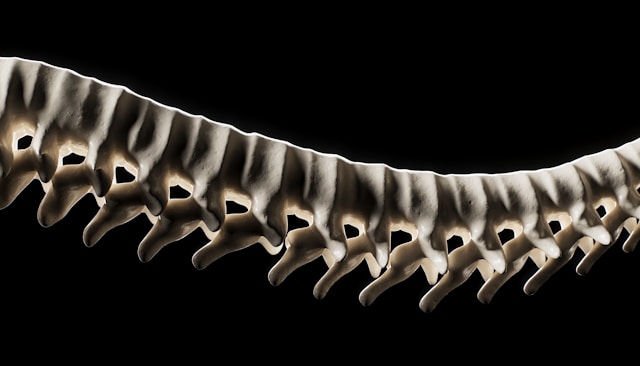
How Imaging Detects Bone Density Changes
The most common and reliable method for detecting early bone loss is dual-energy X-ray absorptiometry (DEXA), also known as bone densitometry. This noninvasive imaging test measures bone mineral density at key sites such as the hip and spine.
Through Bone Densitometry, GLMI provides precise bone density evaluation for patients throughout Buffalo and Western New York.
The test works by using low-dose X-rays to measure how much calcium and mineral content are present in bone. The results are reported as T-scores, which compare an individual’s bone density to that of a healthy young adult reference population.
Even small declines in bone density can be detected long before fractures occur.
Why Waiting for a Fracture Is Risky
Many individuals do not consider bone health until after a fracture happens. However, fractures after age 50 — especially in the hip, spine, or wrist — often indicate underlying osteoporosis.
Hip fractures can significantly affect independence and mobility. Vertebral fractures may cause chronic pain, height loss, and posture changes. Preventing the first fracture is far more effective than treating its consequences.
In Buffalo and surrounding WNY communities, winter falls are common. Early detection of bone weakness can help reduce the severity of injury should a fall occur.
Who Should Consider Bone Density Screening?
Bone densitometry is often recommended for:
- Women age 65 and older
- Postmenopausal women under 65 with risk factors
- Men age 70 and older
- Adults over 50 with a history of fracture
- Individuals with family history of osteoporosis
- Patients on long-term corticosteroid therapy
However, screening decisions should always be made in consultation with a healthcare provider based on individual risk factors.
Risk Factors for Accelerated Bone Loss
Several factors can contribute to faster bone density decline:
- Hormonal changes (especially menopause)
- Low calcium or vitamin D intake
- Sedentary lifestyle
- Smoking
- Excess alcohol use
- Certain chronic medical conditions
In Western New York, limited winter sunlight exposure may influence vitamin D levels, which are important for calcium absorption and bone health.
How Early Detection Changes the Outcome
When bone loss is identified early, physicians can implement strategies to slow progression and reduce fracture risk. These may include:
- Weight-bearing exercise programs
- Calcium and vitamin D optimization
- Fall-prevention strategies
- Medication when clinically appropriate
Monitoring bone density over time allows physicians to track changes and adjust management as needed.
Without imaging, these decisions would rely on guesswork rather than objective data.
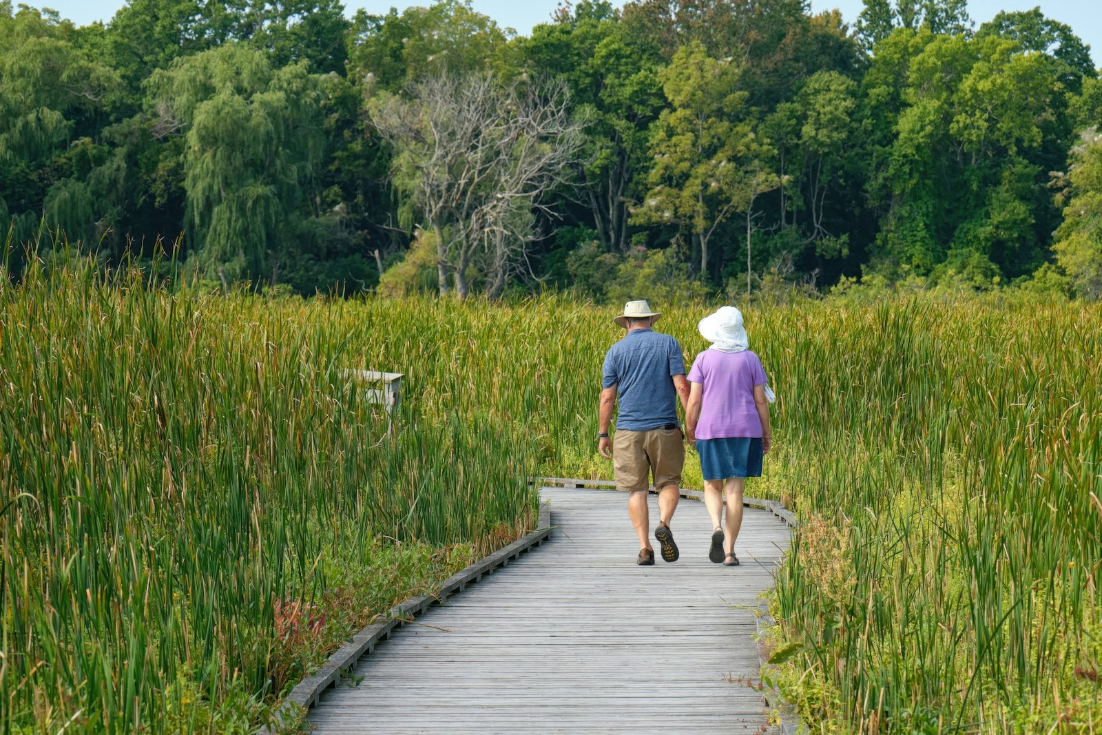
Bone Health and Long-Term Mobility in Western New York
Buffalo and Western New York residents value independence and active living. Whether it’s gardening in spring, walking local trails in summer, or navigating snowy sidewalks in winter, strong bones are essential for maintaining quality of life.
Early detection of bone loss supports continued activity and reduces the likelihood of fractures that could disrupt mobility.
Through comprehensive Imaging Services, GLMI supports preventive health planning across multiple areas — including bone health.
Accessible Screening Close to Home
Convenient access to diagnostic services makes preventive care more achievable. GLMI serves patients throughout Buffalo, Williamsville, Cheektowaga, Orchard Park, and neighboring WNY communities.
With multiple Locations across Western New York, bone density testing can be completed efficiently and close to home.
If you and your healthcare provider determine that bone densitometry is appropriate, you can schedule an appointment here.
Prevention Is Stronger Than Recovery
Fractures often feel sudden, but bone loss is gradual. Imaging allows physicians to detect that gradual change early — before a fall leads to injury.
For adults in Buffalo and Western New York, proactive bone health evaluation represents an investment in long-term independence. Early detection provides clarity, guides preventive strategies, and reduces the risk of unexpected fractures.
Strong bones support active living. Imaging helps protect them — before fractures happen.
Medical Disclaimer: This article is intended for educational purposes only and does not constitute medical advice. Always consult your physician or qualified healthcare provider regarding any questions you may have about a medical condition or appropriate diagnostic testing. Individual health circumstances vary, and decisions about imaging or treatment should be made in consultation with your healthcare provider.
‹ Back
Recent Posts
-
-

-

-

How Ultrasound Helps Diagnose Soft-Tissue Knee Injuries
June 17, 2026
-