
Website by ![]()
Our Blog
The Importance of Radiology in Breast Cancer Treatment
Breast cancer occurs in the breast because of cell mutations in genes that regulate cells’ growth and overall health. When things get out of balance, the abnormal cells attack other healthy cells, causing them to die. One sign that something may be wrong is the feeling of a lump in the breast when performing a self-check exam at home. In other instances, malignant tumors are detected at a radiology screening.
Studies show that around 13% of women will have breast cancer at some point. Men can also develop the disease, though the risk is lower. Radiology is an essential tool in helping physicians and support staff detect and treat patients with breast cancer.
How Can Radiology Detect Breast Cancer?
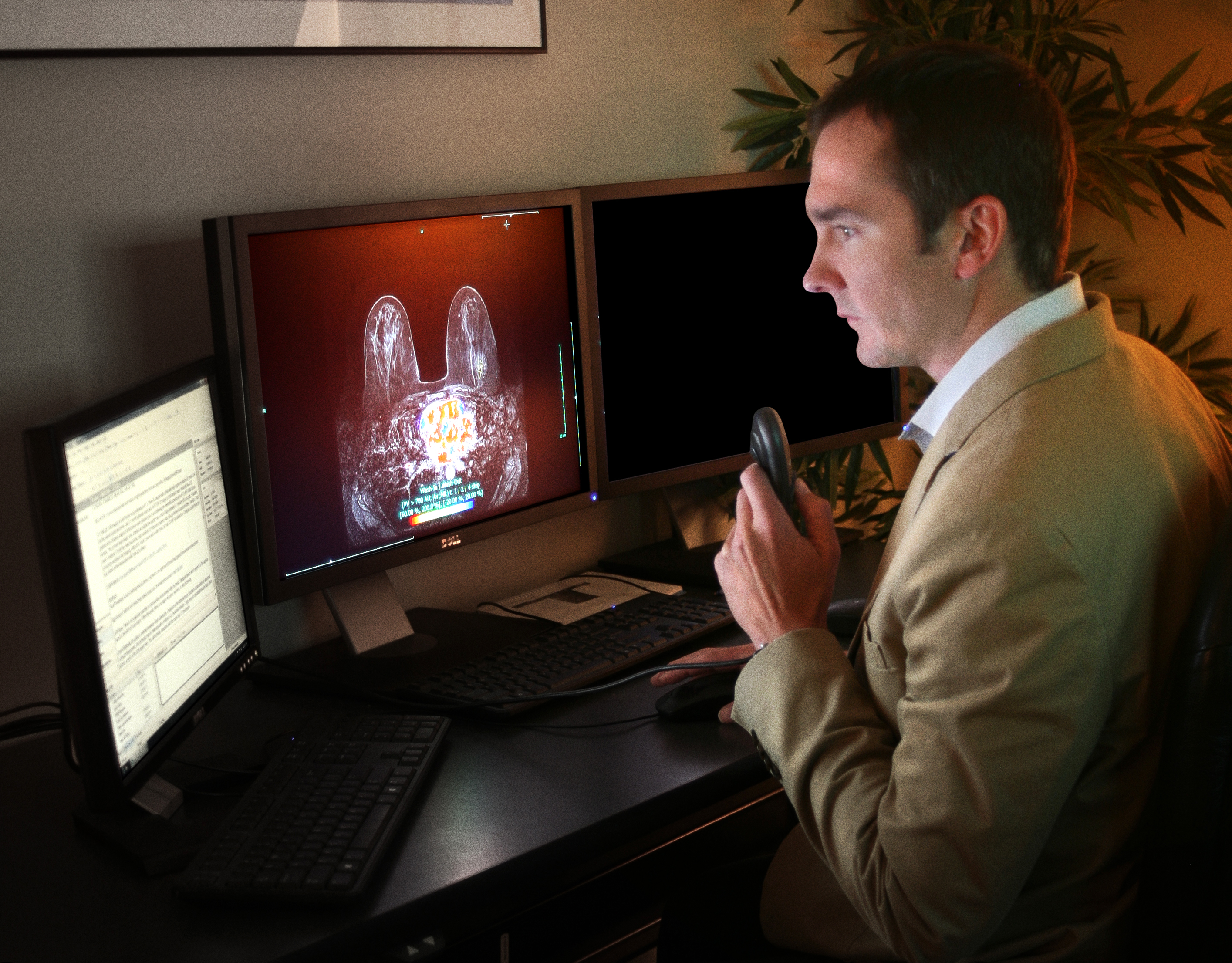
Radiology plays a vital role in diagnosing and tracking the progression or regression of breast cancer during a patient’s treatment. Imaging tests allow doctors to view what’s happening inside a patient’s body. They work by sending some form of energy, like an x-ray or soundwave, through the body.
As those waves travel, the body tissues turn the energy into a pattern that technology can capture and render as a picture or image. They let doctors get a glimpse of what’s happening within the body. That way, health providers can come up with a treatment path to address any issues present, like breast cancer.
Doctors use radiology tests to determine if there is possible breast cancer present. In addition to radiology, physicians take tissue samples and examine them under a microscope to check for any malignancies.
What Radiology Tests Are Used During Breast Cancer Treatment?
Once doctors confirm the presence of breast cancer, they usually order additional radiology testing to determine the extent of its spread. If it’s localized, the tumor is confined to the breast area. If a patient’s breast cancer has spread to the lymph noted, it’s referred to as regional.
Distant breast cancer, also called metastatic breast cancer, is cancer that’s moved to other parts of the body like the bones, lungs, or brain. In those instances, doctors may order special radiology tests to determine how far the breast cancer has spread.
Let’s look at the different types of radiology and how they get used during a patient’s breast cancer treatment.
Mammogram
A mammogram uses low-dose radiology to take a picture of a breast to see if a patient has early signs of breast cancer. They’re capable of finding the disease up to three years before someone might detect the presence of a lump.
They work by having the patient stand in front of a mammogram machine. A technologist places the patient’s breasts on a plate while a separate plate presses down from above. Both plates come together to take an image of the breast. The process is repeated to take a side view of the breasts. Patients typically receive the results back the same day.
Breast Ultrasound
A breast ultrasound image, or breast sonogram, uses sound waves instead of x-rays to get pictures of the inside of a patient’s breast. The radiology test involves placing gel on the skin, then using a small probe to send sound waves into the breast and collect those that come back.
Those waves get sent back to a computer that uses the sound waves to form an image. One benefit of ordering a breast ultrasound radiology test is that they capture more precise images of body organs, including blood moving through vessels. That information can help provide additional information about what’s causing the presence of a mass in the breast.
Breast MRI
A magnetic resonance imaging (MRI) test performed is typically ordered by a doctor after they confirm the presence of breast cancer. It gives physicians more information about the extent of a patient’s breast cancer. In addition, a doctor may recommend that a woman have a breast MRI if they are determined to be at high risk for developing the disease. A breast MRI can provide physicians with multiple images of a patient’s breast.
Computed Tomography (CT) Scan
CT scans allow doctors to create multiple images of cross-sections of the body. They can put them together to create a picture of a specific body part. During breast cancer treatment, physicians use CT scans to determine if any cancer has spread to other body organs like the liver or pancreas.
Is Breast Cancer Radiology Safe?
Radiology tests like a mammogram typically use low doses of radiation. Patients who have questions about the technology behind specific radiology tests should bring any concerns to their physician or the attending technologist. They can answer any questions about the relative safety of the procedure.
The minimal risks involved with using certain radiology tests are outweighed by the benefits they provide. Doctors would not have the tools necessary to track breast cancer and create effective treatment plans for patients without radiology.
Sources
https://www.breastcancer.org/facts-statistics
https://www.cdc.gov/cancer/breast/basic_info/mammograms.htm
https://www.radiologyinfo.org/en/info/breastus
https://www.mayoclinic.org/tests-procedures/breast-mri/about/pac-20384809
‹ Back
Recent Posts
-
-

-

-

-

How Ultrasound Helps Diagnose Soft-Tissue Knee Injuries
June 17, 2026